
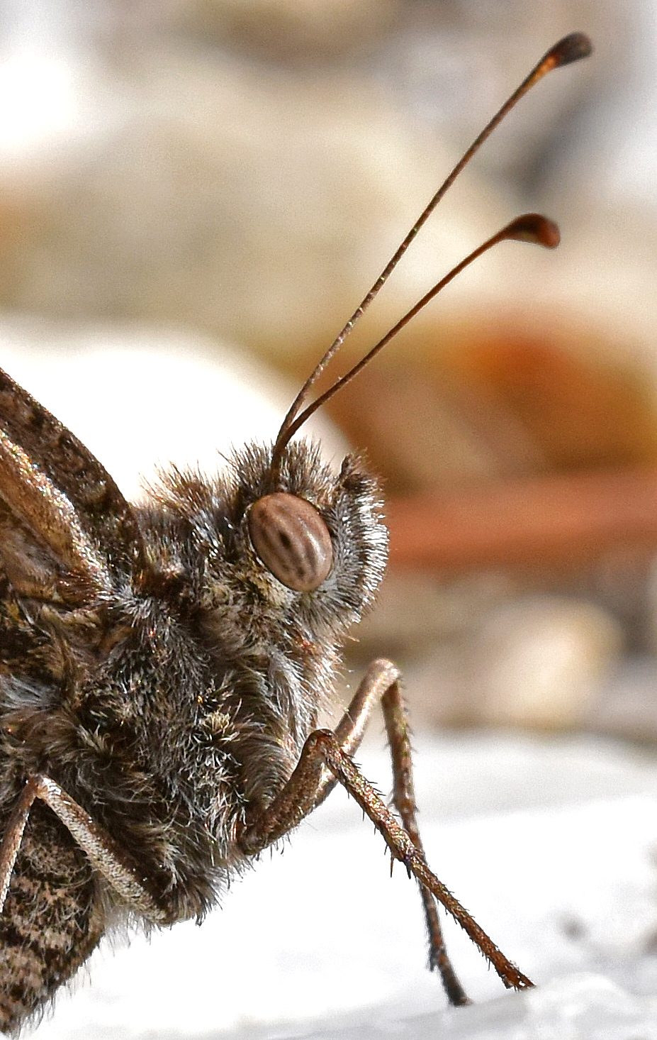
When taking pictures of butterflies, especially when using a flash-gun or pop-up flash, a regular pattern of dark spots often appears in the compound eye. The dark spot near the centre of the eye, as well as the less well-defined (‘accessory’) spots scattered around about it, are called pseudopupils. The largest and darkest spot – the principal pseudopupil – is that part of the compound eye which is facing directly towards the camera. These pseudopupils are not fixed, but move with the angle of view of the observer, or lens (Glaeser & Paulus, 2015).
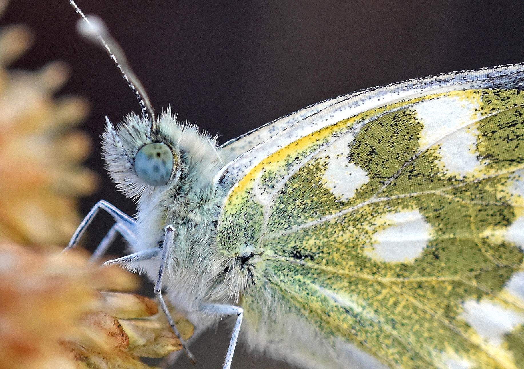
The individual light gathering elements in a butterfly eye are called ommatidia. There can be up to 12,000 of these identical ommatidia, for example in the eye of a swallowtail butterfly; a mosaic of corneal facet lenses. A facet lens sits on top of each ommatidium and immediately below the lens is a crystalline cone. The facets in insect compound eyes are tiny, approx. 15 to 30 micrometers in diameter, yet clearly visibly in a close-up photograph of a butterfly eye. Within each ommatidia there are nine photoreceptor cells, which together form a long, cylindrical, optical-sensing structure called the rhabdom. There are different types of ommatidia, containing different combinations of photoreceptor cells, the light-absorbing visual pigments.
Optics is not an easy subject for beginners (like me!). But essentially, if I have understood it correctly, the principal pseudopupil appears as dark spot because of the black – usually jet-black – pigments clustered closely around the cones. In the words of Professor D. G. Stavenga, of the University of Groningen, NL (pers. comm.), who authored an 80-page publication on this subject, ‘the pigments in the pigment cells surrounding the photoreceptor set of each ommatidium function as shields for stray light … they are usually blackish, absorbing throughout the whole wavelength range.’ So, in the principal pseudopupil, one is in effect looking right down a small group of ommatidia – down their vertical axis – and there is little or no light scattered back by the black primary pigments. In the principal pseudopupil, the black pigment is seen directly through the facet lens of the ommatidium, whereas in the accessory pseudopupils, the pigment is observed through the facet lens of the neighbouring ommatidia (Stavenga, 1979).
According to Professor Stavenga, the compound eyes of butterflies have the richest patterns of pseudopupils. Remarkably, it seems that pseudopupils may have been subject to the same selective forces as the rest of the butterfly body, and the colouration of the eyes could have evolved to fit in with the camouflage or display functions of the wings! This would seem to be a topic ripe for more research; and why not include proboscis colour as well (which often seems to be highly colourful and apparent!).

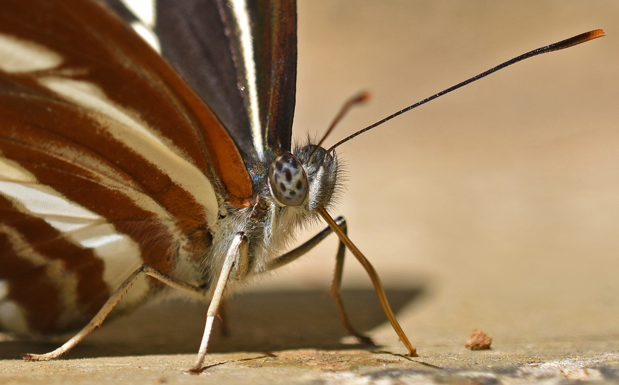
So, in effect, the dark principal pseudopupil is caused by the light being completely absorbed when looking down into the ommatidia, causing those regions to look black (Glaeser & Paulus, 2015). We are looking right down into the photoreceptors and little or no light is reflected back. The main, or central pseudopupil is usually the biggest and darkest one, but in some butterflies (or photographic situations) all of the pseudo-pupils can appear dark, as in the following shot (below).
The shapes and patterns of pseudo-pupils varies to some extent in different groups of butterflies (see Glassberg, 2005).
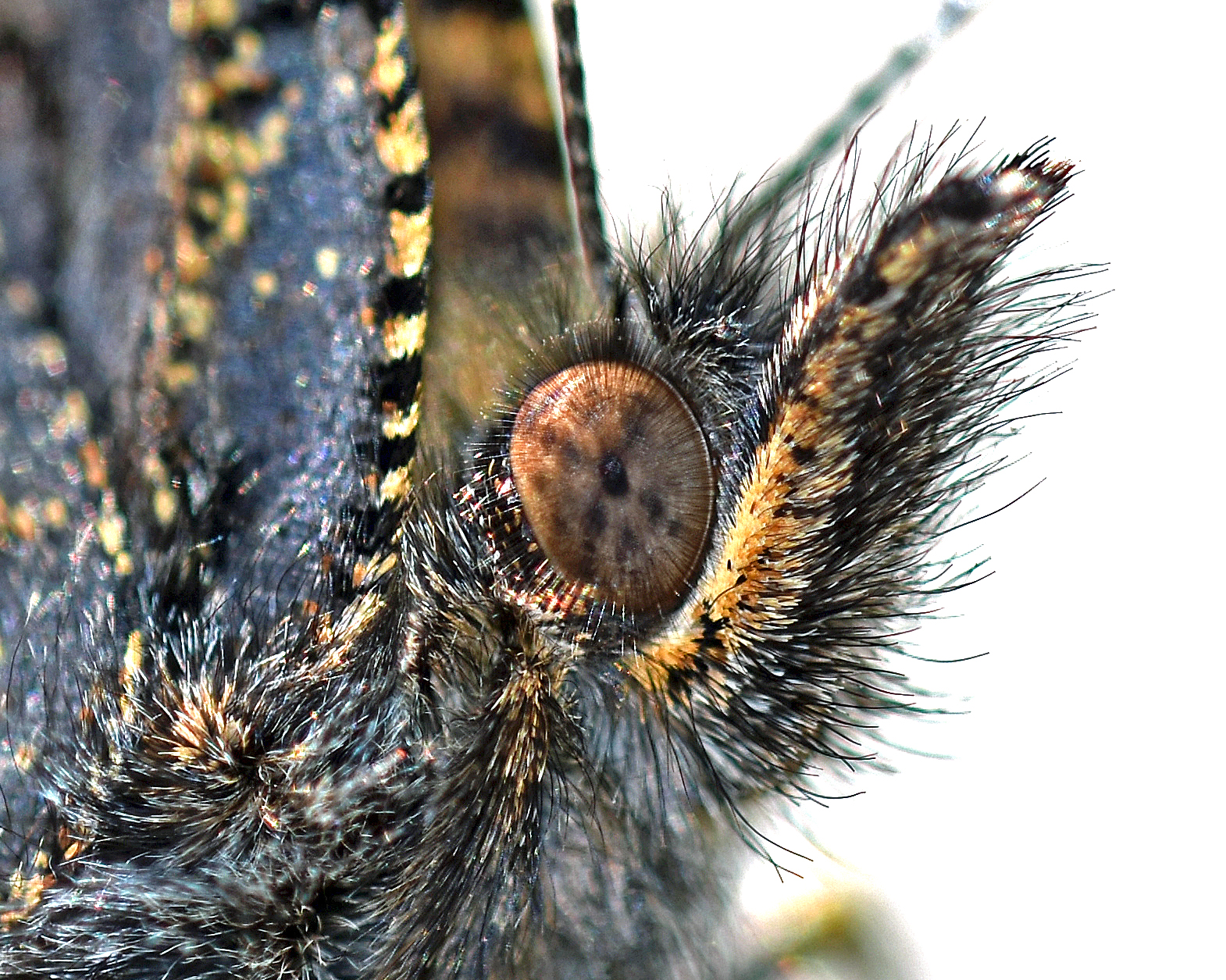
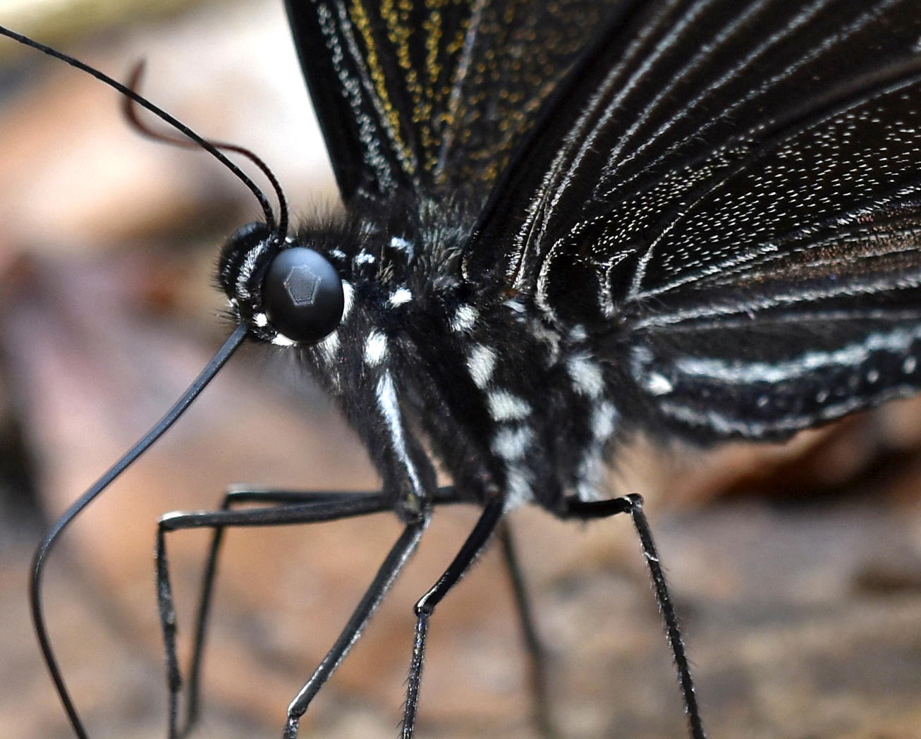
Pseudopupils are said to be ‘optical revelations of the interior of compound eyes’, providing deep insights, into its structure and function (Stavenga, 1979). There is a lot more to it than I have touched on in this blog, and I will finish with a photograph taken of a swallowtail butterfly with large black eyes (below). I used the pop-up flash on my camera to get this shot and I was surprised to see the remarkable hexagonal pattern that appears on the surface of the compound eye (below). It is surely no coincidence that the facet mosaic found in butterflies is hexagonal? Somehow the tiny little hexagonal shaped facets in the compound eye have been magnified up to form a large reflected hexagonal shape, which covers the central part of the eye! That’s optics for you!
I might try butterfly eye colour in another blog!
References
Glaeser, G., & Paulus, H. F. (2015). The evolution of the eye. Springer.
Glassberg, J. (2005). Butterfly Eye-dentification. American Butterflies, 13(2), 34-35. Summer 2005.
Stavenga, D. G. (1979). Pseudopupils of compound eyes. In Comparative physiology and evolution of vision in invertebrates (pp. 357-439). Springer, Berlin, Heidelberg.

Interesting display of butterfly eyes! I’m surprised at the results from pop-up flash, was it adapted in any way?
No, just the little on camera flash.😁
Another interesting Post. Thanks!
[…] the inner structure. They can even be used to look for differences among ommatidia (Stavenga 2002). This blog post gives the best and most accesible summary about pseudopupils I’ve ever […]
Regarding the hexagonal reflection you mentioned at the end – you might find these explanations of their optics helpful (if not to technical): https://farbeinf.de/static_html/strange2.html#fly
https://farbeinf.de/static_html/strange.html#libellen
Thank you.😊
[…] pseudopupils are not fixed, but move with the angle of view of the observer, or lens, as explained here in a blog I did about about butterfly eyes. The pseudopupil is located in the lower sector of the […]